Add us as a preferred source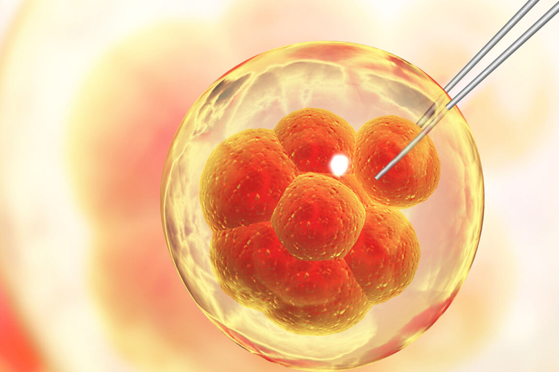
This month’s medical research from British universities – from precision genome editing and dementia research to targeted cancer therapies.
Precision editing to study human embryo development
Researchers led by the University of Cambridge Loke Centre for Trophoblast Research have, for the first time, used an ultra-precise genome editing technique called base editing to study gene function in human embryos.
The study revealed that a specific master gene called NANOG is essential for forming the future body from an early-stage embryo. While base editing has been previously reported in human cells, this project marks the first time the method has been deployed to actively investigate early human embryonic development.
The technique represents a major advancement on conventional CRISPR/Cas9 systems, which can trigger unintended chromosomal abnormalities. Base editing allows scientists to alter a single nucleotide base pair within a genome of approximately 3 billion base pairs. By blocking NANOG, researchers discovered that early embryonic cells failed to develop into specialised pluripotent cells – the critical building blocks that later form the body. Interestingly, the study showed that NANOG functions differently in humans than in mice, where its absence also disrupts yolk sac formation, highlighting the necessity of direct human developmental research.
The breakthrough could eventually help clinical teams improve IVF success rates and better understand early pregnancy loss. It also holds long-term potential for correcting debilitating inherited conditions like cystic fibrosis, though such clinical applications remain legally impermissible in the UK. The unused embryos used in the study were donated by couples who had completed fertility treatment, and research was conducted under strict Human Fertilisation and Embryology Authority regulatory oversight.
“Base editing represents a significant advance on conventional CRISPR/Cas9 because it carries a far lower risk of causing unintended chromosome errors. Base editing can precisely change a single nucleotide base pair to another in an entire human genome of around 3 billion base pairs – that’s an incredible feat,” said Kathy Niakan, director of the Cambridge Loke Centre for Trophoblast Research, who led the study.
CAR T cell therapy targets key mutation driving rare blood cancers
Researchers from UCL and the University of Oxford have demonstrated that a specialised form of CAR T cell therapy can identify and eliminate the cancer-driving stem cells responsible for a rare group of blood cancers, leaving healthy cells unharmed.
The findings target myeloproliferative neoplasms (MPNs) – chronic blood cancers triggered by mutations in blood stem cells.
In roughly one in five cases, MPNs progress to an aggressive, leukaemia-like phase with very limited survival rates and no curative options for the majority of patients. While current treatments can temporarily slow the disease, they fail to eradicate the underlying cancer stem cells. Bone marrow transplants offer a potential cure but are only suitable for a small minority and carry up to a 40% treatment-related mortality risk. To address this, the new therapeutic approach reprogrammes a patient’s immune cells to recognise a specific mutation known as CALR, which is present in approximately one-third of MPN patients.
Using lab-grown human bone marrow models and mice, the research team proved that engineered CAR T cells could selectively kill CALR-mutated cancer cells, even penetrating heavily scarred fibrotic tissue. The researchers also found that combining the therapy with eltrombopag – an existing clinical drug – further enhanced the immune cells’ ability to target the mutation. A Phase I clinical trial is currently being planned at University College London Hospitals (UCLH) and could begin within the next two years, subject to regulatory approval.
“Our model is designed to recreate the real conditions these cancers grow in, including the fibrosis and complex tissue structure that can make treatments fail in the lab-to-clinic gap. Seeing the CAR T cells find and kill the cancer cells in fibrotic marrow organoids was a highly encouraging step,” said Beth Psaila, a senior author from the MRC Weatherall Institute of Molecular Medicine at the University of Oxford.

New targets for dementia
Researchers from King’s College London, working in collaboration with the UK Dementia Research Institute, have discovered a completely new mechanism for brain cell death in patients with Alzheimer’s disease and frontotemporal dementia (FTD).
The process, named karyoptosis, is triggered by the toxic accumulation of proteins inside neurons. When a cell dies via this pathway, the nucleus – the central command zone containing genetic information – shrivels and disintegrates. While other forms of cellular death are well documented, they fail to account for the total neuronal loss seen in neurodegenerative conditions.
The study used advanced computational algorithms to screen 3,000 individual cells from the brains of 28 patients with FTD or terminal-stage Alzheimer’s disease. The data revealed that 35% of cells from the frontal cortex of Alzheimer’s patients exhibited distinct markers of karyoptosis, compared to only 15% in healthy aged controls. The researchers established that toxic protein build-up destabilises the outer structure of the nucleus, coordinating the cascading chemical events that lead to its destruction.
The team successfully reduced karyoptosis markers in laboratory models by targeting the proteins that act as switches in this pathway. Specifically, blocking the interaction between a kinase called p38 MAP kinase and a protein known as LaminB1 slowed nuclear disintegration. This discovery opens up entirely new therapeutic pathways for halting or slowing down brain cell loss, potentially extending the window of opportunity for treatments addressing the root causes of dementia.
“By specifically targeting the interaction between p38 MAP kinase and LaminB1, we may slow down the process of cell death, buying time for more pinpointed therapies against specific neurodegenerative diseases,” noted Manolis Fanto, reader in functional genomics at King’s College London.
Rare ageing disorder links biological clock to disease
An international research team led by the University of Edinburgh has discovered a rare genetic condition that causes accelerated cellular ageing, offering the first definitive proof that the body’s internal biological clock directly drives age-related pathology.
The study settles a long-standing scientific debate by demonstrating that cellular clocks actively contribute to tissue decline rather than simply serving as a passive measure of time.
The breakthrough centred on the identification of Heyn-Sproul-Jackson syndrome (HESJAS), an exceptionally rare disorder in which epigenetic markers – specifically chemical alterations known as DNA methylation – accumulate at the exact same genomic locations seen in normal ageing, but at a vastly accelerated velocity. Patients afflicted with HESJAS presented with classic geriatric health complications much earlier in life, including hair loss, severe osteoporosis, and decreased blood cell production, the latter of which leaves individuals highly vulnerable to recurrent infections.
To understand the underlying mechanisms, researchers deployed a mouse model of the syndrome. They observed that as the premature DNA methylation marks accumulated, adult stem cells – which are fundamentally crucial for ongoing tissue repair and renewal – ceased to function properly. This failure in cellular regeneration triggered metabolic complications closely mirrored by diabetes and elevated cholesterol levels, providing clinical teams with a clear roadmap of how cellular degradation leads to systemic disease. Further global trials are planned to evaluate whether these specific epigenetic shifts can be therapeutically reversed to support future rejuvenation medicine.
“It has been exciting to be able to discover a rare human genetic disorder that helps us understand this clock’s role for all our long-term health in old age. This study has only been possible because of very many generous, often co-located colleagues, who contributed their diverse skills to this work,” said Andrew Jackson, study lead from the Institute of Genetics and Cancer at the University of Edinburgh.
Molecular mapping of adenomyosis lesions points to non-surgical fertility therapies
Researchers at the University of Liverpool have mapped the distinctive molecular profile of adenomyosis lesions for the first time, paving the way for targeted, fertility-preserving treatments.
The study offers fresh insights into a poorly understood gynaecological condition that affects up to 20% of women of reproductive age. Adenomyosis occurs when tissue resembling the womb lining grows deep into the uterine muscle, causing severe pelvic pain, heavy menstrual bleeding, and subfertility.
Historically, therapeutic choices have been highly restrictive, limited to hormone-based regimens that prevent pregnancy or invasive surgery to remove the womb entirely. To overcome longstanding gaps in disease pathology, the research group used spatial transcriptomics – an advanced imaging technique that allows scientists to analyse gene activity within specific cell types while keeping the tissue’s physical structure intact. The analysis revealed that the diseased tissue possesses a distinct biological fingerprint, sharing strong similarities with the deeper, more stable basalis layer of the womb lining.
Crucially, the team identified active indicators of localised inflammation and disrupted cellular energy production within the lesions, which likely drive the debilitating physical symptoms reported by patients. By screening these unique molecular profiles against digital drug libraries, the researchers successfully identified a selection of existing compounds and emerging medicines capable of reversing the disease’s transcriptomic signature. While clinical testing is required, the discovery represents a major milestone toward non-surgical, lesion-specific interventions.
“By identifying the features that distinguish diseased tissue from healthy tissue, we are laying the groundwork for treatments that could directly target the condition while preserving the uterus and normal womb lining. Ultimately, our goal is to develop safe and effective therapies that help women avoid major surgery and protect their fertility where possible,” said Alison Maclean, NIHR clinical lecturer at the University of Liverpool.

Digital diabetes tool shown to improve health and cut NHS costs
A study by researchers at the University of Manchester has linked an NHS-supported digital self-management platform to improved blood glucose control, better overall health metrics, and long-term cost savings for the healthcare system.
The findings evaluated the real-world deployment of the MyWay Diabetes online platform and smartphone app, which allows patients with Type 2 diabetes to track their clinical results, view primary care records and access tailored educational advice.
The analysis followed 507 active platform users for up to two years during the COVID-19 pandemic, comparing their outcomes against a large control group of more than 10,000 similar patients who did not use the tool. The data revealed that MyWay Diabetes users achieved clinically significant reductions in their blood sugar levels alongside distinct decreases in both systolic blood pressure and cholesterol. Crucially, the research team’s economic modelling indicated that the digital intervention is highly cost-effective, offering a scalable mechanism to lower overall NHS spending by preventing future health complications.
Unlike standard standalone health applications, the platform interfaces directly with existing primary care databases to give patients a centralised view of their medical history. Health economists involved in the project emphasised that even minor population-level improvements in cardiovascular risk factors can lead to meaningful reductions in chronic disease burden, proving highly advantageous for financially constrained networks managing large volumes of long-term conditions.
“Managing type 2 diabetes requires people to make complex day-to-day decisions about their health. Our findings suggest that accessible digital tools such as MyWay Diabetes can support people in improving important risk factors linked to long-term diabetes complications,” said lead author Rathi Ravindrarajah from the University of Manchester.
Table sugar and vinegar breakthrough slashes manufacturing barriers for vital drugs
An international collaboration has developed a method for manufacturing carbohydrate-based medicines using table sugar and vinegar.
The breakthrough, co-led by scientists at the University of Bristol and Scripps Research in the US, could significantly lower production costs for critical therapies used to treat Type 2 diabetes, heart failure and chronic kidney disease. The study introduces a simple technique to forge highly stable carbon-carbon bonds that have historically been slow, hazardous and expensive to manufacture.
The research focuses on a vital class of sugar-based molecules known as C-glycosides, which are modified to remain resilient against metabolic breakdown in the human body. To demonstrate the industrial scalability of the process, the team successfully synthesised all currently approved SGLT2 inhibitors – a class of blockbusters including dapagliflozin, canagliflozin, and empagliflozin, which hold a global market value exceeding £15 billion a year. The streamlined chemistry bypasses traditional engineering barriers by completely eliminating the need for complex protective layering, photochemistry, or toxic metal salts.
By mixing standard sugar molecules with a common reagent in mild acetic acid, the researchers directly converted the raw material into a sulfonyl hydrazide that crystallises efficiently. This single-step method sets up the sugar to react as a highly predictable radical precursor, allowing the reaction to be performed easily in standard laboratories worldwide. Because the method has not been patented, the co-authors have openly invited generic pharmaceutical manufacturers to adopt the chemistry to help bring down long-term medication costs for patients globally.
“This discovery could be a total game-changer for manufacturing key medicines faster and more cost-effectively. Due to its operational simplicity and ready availability of the starting materials, I have no doubt it will be the method of choice to make these important molecules in the future,” said study co-lead author Varinder Aggarwal, Alfred Capper Pass chair of chemistry at the University of Bristol.

DNA discovery could shield key healthy cells from chemotherapy side effects
A cellular discovery led by the University of Sheffield, in collaboration with the UT Southwestern Medical Centre in the US, could pave the way for cancer therapies that aggressively target tumours while shielding healthy tissue from debilitating side effects.
The in vitro study identifies a specific pathway to protect irreplaceable, non-dividing cells in the brain and heart during chemotherapy, offering a promising new avenue for oncology research focused on preserving long-term quality of life.
The research focused on a common class of chemotherapies known as TOP2 poisons, which trap the enzymes responsible for cutting and re-stitching DNA during cell division. While these drugs cause cancer cells to self-destruct, they also inadvertently destroy healthy brain and heart cells. However, the team successfully exploited a key biological difference: cancer cells rely on two variations of the enzyme – TOP2A and TOP2B – whereas healthy cells rely exclusively on TOP2B. Using Atomic Force Microscopy, Sheffield scientists observed that a stress-response protein named HSF1 enhances the binding of TOP2B to DNA without interacting with TOP2A. By introducing an HSF1 inhibitor alongside standard chemotherapy, the researchers successfully shielded healthy cells while allowing the drug to retain its full tumour-destroying capacity.
The breakthrough allows clinical researchers to begin testing combination therapies to confirm whether secondary toxicities can be completely prevented in living models. By identifying how these structural regulators function at the genomic level, scientists hope to develop more refined, targeted treatment protocols.
“It is incredible to be able to see how these proteins cling to DNA with nanometre precision. Our advanced imaging methods and analysis techniques are allowing us to begin to understand how the many different proteins in our cells interact with each other and the genome,” said Thomas Catley, postdoctoral research associate at the University of Sheffield.