In the second of a two-part interview, Amanda Randles, director of the Duke Center for Computational and Digital Health Innovation, explains that the shift toward true digital twins is imminent.
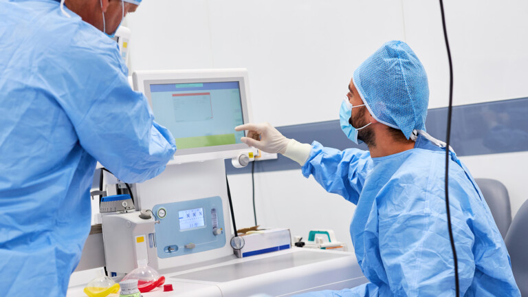
In the second part of her interview with Healthcare Today, Amanda Randles, director of the Duke Center for Computational and Digital Health Innovation, talks about getting the data right, the challenges in validating her model, and that high-level computational medicine is likely to be integrated into everyday life within a decade.
Forgive the cynical question. Are we collecting the right data today – or the wrong data at scale?
Our work currently involves a strategic mix; we are attempting to identify exactly when high-resolution data is essential and when a more streamlined approach will suffice. On one hand, we are pushing forward with projects that rely on standard commercial wearables. The goal here is to ensure the technology is as translatable and accessible to as many people as possible. On the other hand, for specific medical studies, we collaborate with research labs to develop custom wearables that provide the exact resolution required for our models.
A significant issue we encounter is that many metrics provided by consumer wearable devices are actually proxies for the specific physiological markers we need. These devices are often not directly measuring cardiac output or stroke volume; instead, they are estimating these figures – and even blood pressure – based on other available signals.
A major part of our research is understanding when these estimates are good enough and identifying the nuances of relying on such proxies. This is precisely where accuracy can falter, as the reliability of a proxy can vary significantly based on a patient’s body type or skin tone. We must determine exactly when we can trust these proxy measurements and, crucially, in which clinical scenarios we must remain cautious.
“Our goal is to provide clinicians with more comprehensive information.”
How do you validate a model that is unique to each patient?
Our work represents a shift from population-level metrics to personalised health monitoring. Rather than asking if a patient has crossed an arbitrary universal threshold, we are looking at the specific change in their own trajectory.
If a patient’s metrics drop by 15%, the starting point is less important than the drop itself; it is that deviation from their personal norm that flags a need for clinical review. This approach is powerful because it can account for consistent device bias – if a sensor has a slight inaccuracy, that bias remains constant from day one to day five, effectively washing out when we focus on the relative change for that specific individual.
For initial medical applications, we are focusing on patients who already require clinical intervention, such as those with heart failure. These individuals are already undergoing CT scans or right-heart catheterisations, providing us with invasive measurements like cardiac output. We use this high-fidelity data to calibrate the digital model specifically to that person, ensuring we are not merely estimating but truly tailoring the simulation to their unique physiology.
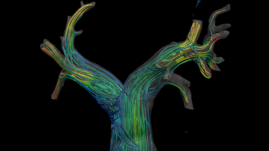
Validation becomes more complex when we move to longitudinal monitoring, as it is historically impossible to measure 3D blood flow continuously over long periods. To address this, we compare our models against data from implantable sensors or discrete daily measurements across a wide range of patients. We also have patients undergo Doppler ultrasound of the carotid artery to obtain specific velocity waveforms, allowing us to verify that our fluid dynamic simulations match real-world observations during both rest and exercise.
Ultimately, a patient’s unique anatomy plays a massive role in how blood flows through their system. A specific heart rate will affect blood flow differently depending on the individual curves and turns of their coronary arteries. Therefore, we make a concerted effort to ensure our models are validated against a diverse range of 3D anatomies. We must ensure the model doesn’t just work for one specific arterial shape; we need to understand exactly how geometry affects the simulation to ensure the model remains robust for every patient.
If these models become widely used, what are the biggest ethical or clinical risks? Who is accountable if a digital twin-informed decision leads to harm?
A crucial point to clarify is that we are not attempting to replace doctors; rather, we are creating clinical decision aids. Our goal is to provide clinicians with more comprehensive information and determine how to present that data in a way that is genuinely helpful.
A doctor does not want, nor could they quickly process, a petabyte of heart rate data spanning 20 years. We are focused on making that information actionable and useful. To that end, we conduct user studies with clinicians to determine the most effective ways to visualise data to ensure they can assess the information accurately and at speed.
In practice, this system acts as a sophisticated monitoring tool that flags anomalies as a patient goes about their daily life. The objective is to alert the doctor so they can make an informed decision ahead of time. In many heart failure cases, for instance, a clinician might be able to intervene remotely by prescribing a statin or adjusting a medication dosage. This improves the quality of care while keeping the doctor firmly in the loop.
Ultimately, we are drawing on the expertise and experience of the clinician to make the final decisions; we are simply providing them with a more complete picture of the patient’s health. That distinction is a critical component of our work.
Doctors are often cynical about innovation. What response are you getting so far?
The medical community has been remarkably supportive, particularly in cardiology. Groundbreaking work by companies such as HeartFlow and CathWorks has already laid the foundation; clinicians have seen the practical utility of these models and are fully on board. The consistent feedback we receive, however, is that they require actionable metrics rather than raw data. A cardiologist does not want a massive zip file of step counts or unfettered access to a patient’s Apple Watch data – they simply do not have the time to sift through it.
Instead, they are excited by tools that can autonomously monitor for specific events or threshold changes and then provide a concise summary. We have been focusing heavily on how to visualise 3D blood flow in a way that is immediately useful. By using augmented reality (AR) and extended reality (XR), we allow clinicians to interact with a patient’s anatomy in an immersive way. This enables them to identify high-risk regions almost instantly.
The response to these tools has been incredible. Clinicians are notoriously time-poor, yet when we run user studies in the cardiology department, they are often so engrossed in the technology that it is difficult to move the study along to the next participant. They recognise that these tools will make their processes significantly faster. We are even refining the simplest details to maximise the accuracy and efficiency with which a doctor can identify a risk region.
The key is to move away from the idea that doctors just want more data; what they truly need is a system that analyses that data for them.
“We are getting remarkably close to a reality where high-level computational medicine is accessible, portable, and integrated into everyday life.”
How close are we to this being usable outside of research environments like universities? What kind of clinical evidence is needed to make that shift?
I believe we will see the first targeted digital twins for specific disease states within the next five years. We are already incredibly close. While we currently have single-time-point personalised models in clinical use, the shift toward true digital twins for specific conditions – such as heart failure – is imminent. Within a five-year window, I expect to see these tools rolling out for a set of defined disease areas, expanding to a much broader range over the next decade.
Furthermore, within the next five to ten years, we may see this technology transition into wellness guidance for the general public, powered by data from personal wearables. This acceleration is largely due to immense advances in computing. The trajectory of GPU (Graphics Processing Unit) computing, combined with breakthroughs in AI algorithms, means we no longer require a massive supercomputer for every single simulation.
These models can now be deployed at the edge. We are reaching a point where these sophisticated simulations can run on an iPhone or a standard mobile device. We are getting remarkably close to a reality where high-level computational medicine is accessible, portable, and integrated into everyday life.