Add us as a preferred source
In the first of a two-part interview, Amanda Randles, director of the Duke Center for Computational and Digital Health Innovation, explains why duration matters.
In the world of modern medicine, the snapshot has long been the gold standard. Whether it is a single CT scan or a discrete blood pressure reading, clinicians have historically been forced to make life-altering decisions based on isolated moments in time. Amanda Randles, director of the Duke Center for Computational and Digital Health Innovation, is working to change that.
A physicist and computer scientist by training, Randles has spent her career at the intersection of high-performance computing and biophysics. Here, she talks to Healthcare Today about the technical hurdles of noisy data, the shift from population-based averages to personalised baselines, and why she wants to replace invasive procedures with non-invasive tools.
Let’s start at the beginning. As you have said, most digital twins today are essentially snapshots. What is the problem with that limitation?
Many of the digital twins we see today are focused on single heartbeats or isolated points in time. The fundamental issue with this approach is that disease is not static; it constantly evolves and changes. To understand a patient’s health, we must capture how the body responds to those changes over time. If we fail to do so, we risk missing the early warning signals and the underlying mechanisms that drive disease progression.
A pertinent example is the identification of vulnerable states. If we only look at a certain snapshot, we are merely asking a binary question: was the patient in a vulnerable state at that specific moment, or were they not? In reality, what matters most is the duration of that vulnerability. Flipping into a vulnerable state for a single second may be medically insignificant; however, if a patient spends three months, or perhaps 10% of their time over a half-year period, in that state, it becomes a serious concern. It is this cumulative exposure that often acts as the driving factor in the progression or localisation of a disease.
When building digital twins for cardiovascular disease, for instance, we often rely on representative heartbeats – such as a single resting beat or a single exercise beat – to understand physiological changes. Yet, a single representative exercise heartbeat fails to capture the different degrees of exertion. It misses how the body responds during the ramp-up to exercise and how it recovers afterwards. It is the data captured across these complete trajectories that is truly telling and, ultimately, predictive.
“This shift towards continuous physiological monitoring is essential.”
You have talked about continuous physiological modelling. What does that mean in practise?
There is currently a continuum on the path toward true digital twins. We have seen personalised models that provide a single snapshot, but I would argue that these do not necessarily constitute a digital twin. To reach that level, one needs constant, continuous feedback from sources such as wearables, sensors, or implantable devices – something that provides real-time data to update the digital model so it accurately reflects the physical entity.
This shift towards continuous physiological monitoring is essential. It allows us to connect wearable data with information from medical imaging, though there is a nuance there: depending on the type of imaging used, those records may also need periodic updates. For example, if a patient has a CT scan but then develops plaque build-up over the following six months, that original scan becomes outdated. We must understand what is happening longitudinally rather than viewing health as a series of discrete events. We need to know when to make those updates and how things are changing.
This approach also grants us the ability to map trajectories and identify shifts from a patient’s own baseline. It makes the process significantly more personalised; we are no longer relying solely on population-based thresholds or arbitrary cut-off points. Instead, we can identify a subtle shift in a specific individual’s average heart rate or recovery time. Even if that shift doesn’t cross a general clinical threshold, the fact that it is a departure from their norm is highly significant. This ability to monitor what is happening for you, as an individual, is where the real power lies.
How hard is this? What are the technical barriers to making that shift right now?
From a technical standpoint, the obstacles are numerous – starting primarily with the data itself. Depending on the clinical focus, we often require incredibly high-resolution, beat-to-beat information. When we integrate physics-based modelling to understand blood flow, we can easily generate terabytes or even petabytes of data if we attempt to capture 3D flow volumes every second over several months. Managing that volume of information is a significant challenge in its own right.
Furthermore, we must contend with the reality of missing or noisy data. At the very least, a wearable device needs to be charged; there will inevitably be periods where a patient leaves their watch on the charger and forgets to put it back on for several days. Then there is the issue of data integrity – for instance, if one of my children picks up my watch and puts it on, how do we identify that the wearable is being worn by the wrong person? We cannot have a system that sends a crisis alert to a doctor simply because my six-year-old is wearing my device.
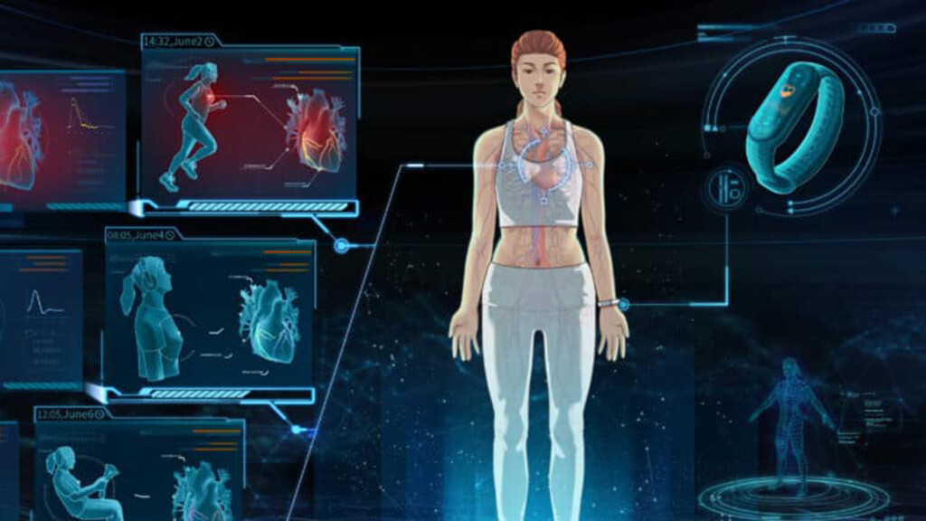
Beyond these practicalities, we currently lack standardised pipelines to aggregate and assess data across different manufacturers and devices. Conventionally, the simulations required for 3D blood flow are computationally expensive. While we are working extensively to train AI surrogates to create reduced-order models, progress varies depending on the specific disease area or biomarker. Integrating these disparate models – combining flow data with heart rate, sleep, gait and even dietary information – presents a massive multimodal data challenge for computer science.
Finally, these technical hurdles do not even touch upon the essential concerns of privacy, security and fairness. Many of the clinical thresholds we use today are based on non-representative populations. Ultimately, we do not just need better models; we require an entire ecosystem to ensure that this technology is feasible, fair and safe as we move forward.
Where can it help? You’ve suggested simulations could complement or even replace invasive measurements. What procedures are most likely to be displaced first?
The most exciting aspect of this field is that we are already seeing these concepts in practice, particularly through what I would categorise as advanced personalised models. A prime example is the measurement of Fractional Flow Reserve (FFR), which is used to determine the burden of coronary ischaemia and whether a patient requires a stent. Conventionally, this involves an invasive procedure where a guide wire is inserted into the coronary arteries to measure pressure. Today, however, companies such as HeartFlow and CathWorks have FDA-approved technologies that are used globally. Instead of an invasive wire, they use a 3D simulation to measure FFR in a virtual replica of the patient. Large-scale clinical trials have already proven that these virtual replicas can match the accuracy and specificity of invasive measures.
Initially, the primary impact of this technology will be in diagnostics. We are starting with coronary arteries, but these methods will soon be applied to cerebral and peripheral vasculature. The goal is to replace invasive procedures with non-invasive tools wherever we have established biomarkers and thresholds. For instance, we are conducting studies on heart failure, where we know that pulmonary artery pressure (PAP) changes before physical symptoms appear. While patients currently require implantable sensors to monitor these changes, our pilot studies have shown that computational models can capture this data non-invasively.
The next step is to monitor these changes over time. If PAP typically changes two weeks before a crisis, can we identify a shift in the trajectory three or four weeks ahead of time by monitoring other biomarkers? Access to this longitudinal data may even allow us to discover entirely new indicators of disease.
Once these computational tools are established, we can use them to project future outcomes. Before a patient enters the operating room, we could virtually try out different stents, bypass procedures, or even the effects of various drugs like statins. We can adjust the virtual patient’s geometry and activity levels to predict the physical forces and long-term effects of a specific treatment. This represents the next wave of progress: using digital twins to inform treatment paths and monitor patients as they go about their daily lives.
“The challenge lies in whether we are capturing the right data.”
Are current consumer devices (Apple Watch, Fitbit) clinically robust enough to feed into models like HARVEY?
This depends entirely on the specific clinical question being asked. In many instances, if we are simply trying to determine whether a patient spent 20% versus 50% of their day in a high-risk state, the data is already sufficient. We do not necessarily require a measurement to the fifth decimal point to understand that a patient has crossed a significant threshold. In those cases, the existing data is more than good enough to inform a model.
However, the challenge lies in whether we are capturing the right data. While some methods provide high-resolution information during periods of exercise, they may only provide an average over an hour for the resting state. If our goal is to capture beat-to-beat variability, that level of averaging is inadequate.
Furthermore, a significant hurdle remains regarding the validation of these physiological signals. Much of the existing validation has been conducted on populations that are not universally representative.
We know, for example, that certain optical sensors may be less accurate depending on an individual’s skin tone. This is an issue that must be addressed with urgency to ensure that the data driving these digital twins is accurate for everyone. While there has been immense progress, the variability between devices and biomarkers means there is still considerable work to be done to ensure these systems are both precise and equitable.